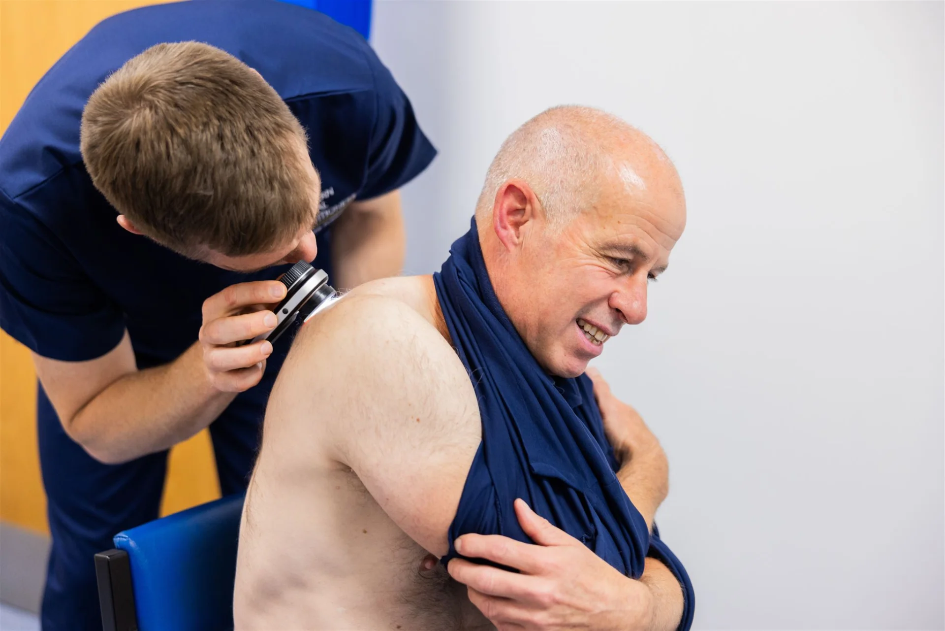
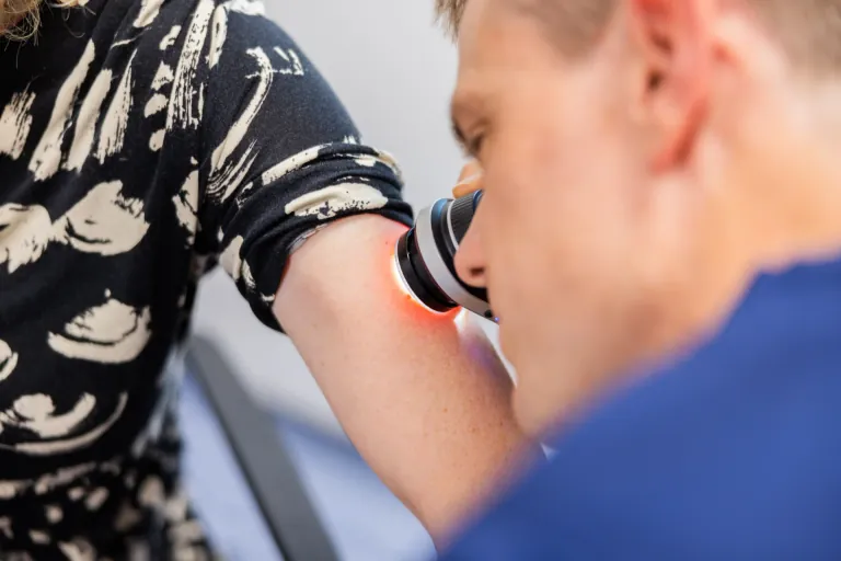
mINOR sURGERIES

Seborrhoeic Keratosis Removal in Newcastle

Seborrhoeic keratoses are very common benign skin growths that often appear with age and are sometimes referred to as “age warts”. They can vary in colour from light brown to black and may look warty, scaly or “stuck on” to the skin.

Although harmless, they are frequently mistaken for moles and can become irritated, catch on clothing, or be cosmetically bothersome.

At Northern Medical Practitioners in Newcastle, we offer assessment and removal of suitable seborrhoeic keratoses in a medical setting, with a focus on safety, clear explanation and good cosmetic outcomes.

Removal of Skin Lesion 1024x683 1

Seborrhoeic keratoses explained

Seborrhoeic keratoses are non-cancerous skin growths that commonly develop in adulthood. They may occur on the face, trunk or limbs and often increase in number over time. While they are benign, their appearance can be alarming, particularly if they darken, thicken or become inflamed.

An experienced clinician can often recognise seborrhoeic keratoses based on their typical appearance and examination.

Why people choose removal

Seborrhoeic keratoses are often removed because they catch, bleed, become inflamed, or cause cosmetic concern. Some patients choose removal because a lesion has changed in appearance or is repeatedly irritated by clothing or jewellery.

We will discuss whether removal is appropriate in your individual case and explain when monitoring or referral to another service is more suitable instead.

How removal is carried out

Removal is usually performed using minor surgical techniques such as curettage or electrodessication, depending on the size and location of the lesion. In cases where confirmation is appropriate, removal can be carried out using a technique that allows tissue to be sent for laboratory (histological) examination.

The most suitable approach will be discussed with you at your appointment.

Aftercare and healing

After removal, the treated area usually heals over 1–2 weeks. You will be given clear aftercare advice to support healing and minimise the risk of infection or scarring. Mild redness or scabbing is common initially.

We will explain what to expect during healing and when to seek advice if recovery does not progress as expected.

Booking Your Seborrhoeic Keratosis Removal

If you are considering removal of a seborrhoeic keratosis, you can book directly for a private appointment at our Blaydon clinic. Many patients are suitable for assessment and treatment in a single visit.

We also offer a free assessment of photographs, which can help confirm whether a lesion appears suitable for private removal and provide an estimated cost where possible. Photo assessment is used to guide next steps and does not replace face-to-face clinical examination.

Price List

Seborrhoeic Keratosis Removal Pricing

Bespoke pricing available for large number of lesions, please enquire.
Initial call
Online Assessment of Photos

Free

Procedure
First Seborrhoeic Keratosis Removal

350

Procedure
Additional Seborrhoeic Keratosis Removal

100

Samples
Histology Analysis of Tissue

105

Aftercare
Follow-up appointment (if required)

Free

Consultation
Face to Face Consultation (no procedure)

100

Frequently Asked Questions

Seborrhoeic Keratosis Removal FAQ's

What is a seborrhoeic keratosis?

A seborrhoeic keratosis is a very common, benign skin growth that tends to appear in adulthood and becomes more common with age. They can vary in colour and texture and are often described as looking “stuck on” to the skin. They are not skin cancer.

No. Seborrhoeic keratoses are different from moles, although they are often mistaken for them. Their appearance can sometimes cause concern, particularly if they darken or thicken, which is why medical assessment is helpful before removal.

Seborrhoeic keratoses are usually removed because they catch on clothing, bleed, become irritated, or cause cosmetic concern. Some patients choose removal for comfort or reassurance rather than ongoing monitoring.

Removal is usually performed using minor surgical techniques such as curettage or electrodessication. The most suitable method depends on the size, thickness and location of the lesion and will be discussed with you at your appointment. Procedures are typically quick and well tolerated.

Seborrhoeic keratoses are benign and do not usually require laboratory testing. However, as a doctor-led service, we are able to use removal techniques that allow tissue to be sent for histological examination when appropriate. This decision is based on clinical judgement and will be discussed with you before treatment.

Seborrhoeic keratoses often have a characteristic appearance, but no assessment is infallible. If a lesion appears atypical or unsuitable for private removal, we will explain this and advise appropriate referral rather than proceeding.

Sending the tissue which has been removed for laboratory analysis (histology) provides a further safeguard.

A treated seborrhoeic keratosis does not usually recur. However, people who develop one seborrhoeic keratosis often develop others over time, sometimes in different areas.

Any procedure that breaks the skin can leave a mark. We will discuss likely cosmetic outcomes beforehand and use appropriate techniques to minimise scarring where possible. Most areas heal with minimal long-term change.

Yes. Multiple seborrhoeic keratoses can often be treated in a single visit. This will be discussed during your consultation, including suitability and pricing.

Yes. We offer a free photo assessment, which can help confirm whether a lesion appears suitable for private removal and provide an estimated cost where possible. Photo assessment is used to guide next steps only and does not provide a diagnosis or exclude skin cancer.

The photo assessment helps us:

  • assess whether the lesion appears suitable for private removal

  • identify whether location or size may affect treatment approach or pricing

  • reduce unnecessary clinic visits

It does not replace face-to-face examination and is not used to diagnose or rule out skin cancer.

To help us give useful guidance, please send:

  • one clear close-up photo of the lesion, showing surface detail

  • one photo taken from further away to show the lesion’s location on the body

If you have more than one lesion, please include photos of each.

For best results:

  • use good natural light where possible

  • ensure the image is sharp and in focus

  • avoid shadows, glare or heavy zoom

  • keep the camera steady

  • ask someone else to take the photo if the area is difficult to reach

Clear photos allow us to give clearer guidance.

No. It is not possible to diagnose or exclude skin cancer from photographs alone. If a lesion appears atypical or concerning based on the images or your description, we will advise appropriate in-person assessment or referral rather than private removal.

Yes. There is no charge for photo assessment or initial guidance. Any fees for consultation or treatment will be explained clearly before booking.